Fabripullus
The Chick of Girolamo Fabrizi
First
part
The formation of the eggs of the birds
Chapter I - Description of the uteri of the bird
The
asterisk * indicates that the item is present in lexicon ![]()
|
HIERONYMI
FABRICII |
GIROLAMO
FABRIZI d’ACQUAPENDENTE |
|
[1]
De |
First
part |
|
Instrumentorum
seminis tractationem proxime formationis foetus tractatio
consequitur. Ea enim lege semen gignitur, ut ex eo foetus procreetur.
Animalium autem foetus, alius ex ovo, alius ex semine, alius ex putri
gignitur; unde alia ovipara, alia vivipara, alia ex putri, seu sponte
naturae nascentia αὐτόματα Graece
dicuntur. Aristot.[1]
aliam formationis foetus speciem ponit, cartilagineorum piscium, qui
partim ovipari, partim vivipari sunt: quia, cum ovum intra se gignant,
foras vivum foetum pariunt. Sed haec diversa a propositis non est, sed
potius mixta, aut ex oviparo, et viviparo composita, ut ostendimus in
libro nostro de formato foetu in picturis. |
The
treatment of the formation of the fetus immediately follows the
treatment of the instruments of the semen. In fact the semen is
produced so that the fetus is procreated from it. Some fetuses of
animals are born from the egg, others from the semen, others from
putrid matter. Thence some animals are called oviparous, others
viviparous, others born from the rotten matter, that is, for a
spontaneous act of nature, in Greek are called autómata -
spontaneous. Aristotle* suggests another type of formation of the
fetus, that of the cartilaginous fishes, partly oviparous, partly
viviparous, for although engendering an egg within themselves, they
give birth outside to a living fetus. But this manner is not different
from the listed ones, on the contrary is mixed, or better, composed by
an oviparous and by a viviparous, as I show in the iconography of my
book on the already formed fetus. |
|
Nos de omni
formatione dicemus. In hac re, ut video, natura imprimis de loco fuit
solicita, quem aut in animali, aut extra animal constituit: atque in
animali uterum esse voluit, extra vero ovum: in utero quidem ex
semine, et sanguine; in ovo vero ex iis, quae in ipso consistunt,
foetus generationem molita est. Quare si intentio nostra est in
praesentia agere de foetus formatione, de utraque agendum est, initio
ab ea, quae ab ovo procedit, desumpto. Haec enim omnem aliam
tractationem praecedere omnino debet: tum quia ex hac Aristo.
opinionis intelligentia non difficulter elicitur, et habetur: tum quia
tractatio formationis foetus ex ovo amplissima est, et altera longe
latior, et difficilior. |
I
shall speak of all the types of generation. On this point, as I
realize, first of all the nature has been attentive in choosing the
place she established to be either inside the animal or outside the
animal, and she wanted that in the animal there was the uterus, but
the egg on its outside. Actually she foretold that the generation of
the fetus in the uterus comes from the semen and from the blood, while
in the egg it comes from the structures present in it. Therefore if at
present it is my intention to treat the formation of the fetus, we
have to treat both the types of formation, starting from that
beginning from the egg. In fact this absolutely has to precede every
other treatment: both because from this the understanding of the
opinion of Aristotle is gathered without difficulty and we acquire it,
and because the treatment of the formation of the fetus from the egg
is very wide, and more wide and difficult than the other. |
|
Amplissimam
autem esse formationis foetus ex ovis contemplationem ex eo patet,
quod maxima animalium pars ex ovis gignitur. Nam ut insecta ferme
omnia, et imperfectiora omittam animalia, quae ex ovo fieri sensui
apparet; ex perfectioribus quoque, maxima pars ex ovis gignitur.
Etenim primum ex volatilibus, omnia pennata, ex aquatilibus vero, si
sola cetacea excipias, caetera omnia, pisces omnes, item crustacea
omnia, et mollia, et testacea ovipara sunt: ex terrestribus, vero sola
nonnulla quadrupedia, et homines vivum foetum edunt; caetera ut
reptilia, multipedia[2],
et serpentia omnia, sunt ovipara, [2] sicuti inter quadrupedia omne
lacertorum genus. Quod si singularia quaedam excipiantur, ea,
enunciationes veras esse, non tollunt. |
Actually
the investigation about the formation of the fetus from the egg is
very wide because the majority of the animals arises from the eggs. In
fact, omitting almost all the insects and the most defective animals
that for perceptible experience come from the egg, also the majority
of those more perfect arises from the eggs. In fact if from flying
animals in first place all the feathered ones are excluded and from
the aquatic ones only the cetaceans, all other animals, all fishes and
likewise all crustaceans lay eggs, both soft and endowed with shell.
Among the terrestrial animals only some quadrupeds and the human
beings give birth to a living fetus; the other animals, as the
reptiles, the myriapods and all the snakes are oviparous, as among the
quadrupeds is oviparous every kind of lizard. But if some single
animals are excluded, they don't remove the fact that the statements
are true. |
|
De Pulli
autem procreatione antequam dicere instituo, prius de ovi generatione
omnino praemittenda disputatio est, quandoquidem in pennato duplex
quodammodo conceptus fit, ovi, et pulli. Utriusque suus uterus est:
pulli quidem ovum; ovi autem quod in pennata faemina, intus proprium
organum positum est, quod ego perpetuo ovarium appellabo; quousque a
probato auctore alio nomine appellatum, inveniam. Ovi hic locus, seu
uterus, seu matrix, duplex est, alius superior, alius inferior.
Superior uterus prope thoracem est ad pennati spinam sub iecore, et pulmonibus
statim, et supra magnam tum venam, tum arteriam[3]. |
However,
before I begin to speak of the generation of the chick, first of all
it is necessary to prefix the treatment about the generation of the
egg, since in a bird a double conception is practically occurring, of
the egg and of the chick. Their uterus exists of both: for the chick
the egg, for the egg that specific organ that in the female of a bird
is set inside and always I will call ovary until I won't find it
otherwise called by a competent author. This seat of the egg, whether
called uterus or matrix, is double, one upper and one lower. The upper
uterus lies close to the thorax near the spine of the bird,
immediately below the liver and the lungs, and over both the great
vein and the great artery. |
|
Arist. 6. de
hist. anim. cap. 2.[4]
dicit ad septum transversum ovum inchoari: nos autem in respirationis
tractatu negavimus pennata septum obtinere. Solvitur dubium, pennata
septo prorsus non destitui, quia membranam[5]
habent tenuem loco septi positam, quam Arist.[6]
cinctum, et septum appellavit:
sed non habent septum, quod musculus sit, et ad
respirationem conferat, ut alia animalia. Aristoteles autem musculum
non agnovit. Inferior uterus sub primo statim ad spinam similiter
ponitur, et utrinque etiam ad lumbos, sed ad podicem usque descendit;
ibique a sinistris finitur. |
Aristotle
in Historia animalium VI,2 says that the egg starts in
proximity of the diaphragm, but in the treatise on respiration I
affirmed that the birds don't have a diaphragm. The doubt is solved:
the birds are not completely devoid of diaphragm, being that they have
a thin membrane there where the diaphragm is, which Aristotle called
cincture and septum; but they don't have, like the other animals, a
septum made up by muscle and useful for the respiration. Actually
Aristotle didn't know a muscle. Likewise the lower uterus is located
below the first one, immediately near the spine as well as near the
loins on both sides, but it goes down until the cloaca and here it
ends on the left side. |
|
Superior
matrix nil aliud est, quam infinita propemodum vitellorum multitudo,
quae in uno veluti acervo conglobata conspicitur, rotundae figurae, et
cuiusvis magnitudinis, in qua a minimo ad maximum ea intercedit
differentia, quae est a grano sinapis ad fructum fere nucis iuglandis,
aut mespili. |
The
superior uterus is nothing but an almost endless multitude of yolks
grouped together as in a single heap, endowed with round shape and of
any size varying from a minimum to a maximum between a grain of
mustard* and approximately a fruit of walnut-tree or of medlar*. |
|
Haec
vitellorum multitudo simul quasi racematim apposita, collecta, et
coniuncta est; ob quam causam ego perpetuo vitellarium, aut vitellorum
racemum appellabo, cum indecorum ut Celsus loquitur[7],
hoc est sine nomine, ab antiquis relictum esse hoc organum videam. Satius
autem forte fuerit vitellarium appellare corpus vitellos expullulans,
ac producens, hoc est vitellorum fundamentum; racemum vero tum corpus
praedictum, tum vitellos racematim appensos, qui Graecis vitellorum βότρυς
dici potest, et nostris quoque botrus. Appello autem hanc partem
racemum, quia uvarum
racemo quam simillima est. Quod et Arist. 3. de gen. an. cap. 8. dixit[8],
cum ait: reddunturque ova eorum glutino cohaerentia ad speciem uvae.
Etenim sicuti in racemo uvae {seu} acini sunt tum maiores, tum minores,
tum minimi, singuli suo pediolo appensi; sic in proposito vitellorum
racemo videre est. |
This
crowd of yolks is placed, gathered and conjoined almost like a cluster,
that's why I will always call it vitellarium or vitellorum
racemus - cluster of yolks, since I notice that this ugly organ,
as Celsus* says, has been left without name by ancients. But perhaps
it would be better to call vitellarium a structure that makes
sprout and produce yolks, that is, the point of origin of the yolks,
and to call racemus -
cluster - both the above-mentioned structure and the yolks arranged as a cluster
which by Greeks can be said bótrys - cluster - of yolks, and
also botrus - grape's cluster - by our fellow countrymen.
Actually I call this part racemus since it is very similar to a
cluster of grape. And Aristotle also told this in De generatione
animalium III,8 when he affirms: their eggs are aggregated by a
sticky substance as being grape. And in fact as in a cluster of grape
the grapes sometimes are greater, sometimes smaller, sometimes
dwarfish, every one suspended to its own stalk, the same it is
possible to see in the described cluster of yolks. |
|
Hic vero
pediolus nil aliud est, quam membranosum corpus, seu nexus robustus
cavatus, qui a racemi fundamento ad vitellum producitur, quem cum
contingit, dilatatur, et perinde ac nervus opticus in oculo
amplificatus, vitellum externa tunica obducit, nec quidem totum
vitellum circundat, sed paulo illum ultra medietatem comprehendit;
perinde ut in glande operculum retro appositum, calix appellatum; quo
fit, ut exterior vitelli portio a proposita membrana destituta
conspectui sese offerat sine venis, et nudata appareat. Huius
membranae exactam quantitatem discernes per vestigium orbiculare, quod
in perfecto vitello visitur, quando membrana suis terminis a vitello
laxari, et resolvi incipit: tunc enim denudati vitelli vestigium quasi
zona vitellum cingens apparet. |
But
this stalk is nothing but a membranous structure or the strong hollow
connection extending from the base of the cluster to the yolk, and it
dilates when reaching it, and as if being the optic nerve that widened
in the eye, it surrounds the yolk with an external tunic, and it
doesn't totally surround the yolk, but it winds it a little more than
half, like the cover called cup does in an acorn, being leant at the
back. Then it happens that the external part of the yolk, without the
described membrane, offers itself to the eyes without veins and
appears naked. You will realize the exact extension of this membrane
by a circular vestige that is observed in a completed yolk when the
membrane begins to grow loose and to free itself from the yolk in
correspondence of its periphery. In fact then a trace of the naked
yolk appears as if being a girdle surrounding the yolk. |
|
Huius
pedunculi, et dimidiatae membranae [3] beneficio increscens vitellus,
quasi suspensus, et caeteris elatior detinetur. Qui sane pedunculus
etiam secum multa vasa in vitellum deducit, quorum maiora per pediolum
discurrentia subinde in vitellum propagantur, ac disseminantur. Porum
hunc, seu fistulosum canalem, qui [vitellarios] vitellarii singulos
coniungit, et suspensos detinet vitellos, perpetuo pediculum, pediolum,
et pedunculum appellabo, quod fructuum pediculo quam simillimus sit;
quem forte Arist. 3. de gen. an. cap. 2.[9]
στόλον
{ὀμφαλώδην}
<ὀμφαλώδη>,
hoc est appendiculam umbilicalem, et veluti fistulam nuncupavit; licet
non ex omni parte descriptio competat, forte propter secundi uteri
ignorationem. |
The
yolk,
increasing thanks to this peduncle and the dimidiate membrane, is
maintained almost suspended and more elevated than the others.
Actually this peduncle also brings with itself many vessels in the
yolk, the larger of them, by flowing through the petiole, subsequently
propagate and spread in the yolk. This passage, or tubular channel,
connecting the single yolks of the cluster and maintaining them
suspended, I will always call pediculus, pediolus and pedunculus,
since it is extremely similar to the petiole of the fruits. Perhaps
Aristotle in De generatione animalium III,2 called it stólon
omphalřdë - navel shaped prominence, that is, umbilical appendix
similar to a tube. Although the description is not fully suitable,
perhaps because of the lack of knowledge of the second uterus. |
|
Vitelli autem
in racemo maiores in circuitu sunt, minores in medio, ceu a maioribus
circundati, denique minimi omnibus subiecti: rursusque minimi duriores,
ac caeteris validiores: ideoque vitelli iustam magnitudinem adepti
mollissimi omnium sunt. Prodeunt hi a suo fundamento, quod est corpus
quoddam sui generis, ex semine a primordiis ad pennati spinam ob ortum,
fixum, obfirmatumque, quod temperie a mediocritate vix recedit, colore
potius albescens; consistentia inter molle est, et durum, sed tamen
laxum et porosum; figura rotunditati aemulum; numero unum; uniforme,
et magnitudine moderatum, sed oblongum, et in summitate amplificatum,
ut plurimos, numerososque vitellos ex se producat, prius singulari
pediolo singulis expullulato. Hoc sane corpus substantiae proprietate
vitellos expullulare, generare, et ex sanguine producere aptum est;
idque non solum privatum, quo se ipsum nutrit, sed etiam publicum
agens munus, speciei propagationi, et conservationi substituitur,
ovoque initium exhibet, et praebet, vitellos ante omnia efformando. |
Besides
in the cluster the yolks are greater in the periphery, are smaller in
the middle part, as surrounded by the greater ones, finally the
dwarfish ones are placed under all of them. Besides the dwarfish ones
are harder and more solid than the others, then the yolks that reached
the correct size are the softest of all of them. These escape from
their base, a quite particular structure, because of the constant and
stable birth from the semen starting from the primordial elements
placed near the spine of the bird, and that for structure just strays
from the right, being of rather whitish colour. For consistence - this
structure - is between the soft and the hard, but nevertheless it is
soft and porous, for the aspect emulates the roundness, is only one in
number, is uniform and of moderate size, but lengthened and magnified
at the summit so that it produces any amount of yolks and numerous
yolks, before for each one the own peduncle is formed. Actually this
structure, for the characteristics of the substance constituting it,
is proper for to bud, to generate and to produce yolks from the blood.
And besides it undertakes the propagation and the maintenance of the
species, not only carrying out a private task thanks to which it
nourishes itself, but also a public one, and it causes the beginning
of the egg and provokes it by moulding the yolks before all the other
things. |
|
Hi vitelli
sicuti initio a parvulo incipiunt, ceu milii, aut sinapis magnitudine,
et minuti sunt ac candidi, ut dicit Arist. 6. de hist. an. cap. 2.[10]
sic subinde paullatim increscunt, et ut ait Arist. lutei ac flavi
efficiuntur, quousque ad iustam magnitudinem omnibus notam perveniant.
Hoc tempore iam a suo pedunculo, ac tota eiusdem membrana,
amplificatione eorum attenuata, separantur, abrupta a pediculo
exporrecta eiusdem membrana, separatione hac ita facta ambo, hoc est
tum pedusculus, tum membrana paulo post contrahuntur, et in suum
fundamentum, nimirum corpus, a quo producta sunt, retrahuntur, eique
associantur atque coniunguntur, quasi in pristinam naturam conversa;
ubi videre interdum est huiusmodi tunicas abruptas tres, quatuorve;
quae uti, dum vitellum involvebant maxime distentae tenuissimaeque
apparebant; ita abruptae, et contractae, atque in proprium corpus
reversae, quasi uterus effoetus, ad pristinam naturalem magnitudinem
redeunt. |
Like
these yolks start from a very small initial structure, of the size of
a corn of mile* or of mustard*, and they are small and white, as
Aristotle says in Historia animalium VI,2, so subsequently they
increase gradually, and, as Aristotle says, they become yellow and
golden, up to reach the correct dimension known to everybody. In this
moment, their growth being reduced, by now they separate from their
pedicle and from all its membrane, its wide membrane having detached
itself from the peduncle, and after this separation is so realized,
they both, that is both the pedicle and the membrane soon after
contract and withdraw in their point of origin, that is, the structure
whence they produced themselves, and they join it and unite to it,
almost they came back to the original shape. Where sometimes it
happens to see three or four of such broken tunics, which until
surrounding the yolk appeared extremely widespread and very thin. So
broken and refold, and turned on their structure as being a
post-partum uterus, they return to the natural primitive size. |
|
Vitellus vero
unica propria, et ea
quidem tenuissima tunica obductus, in membranosam, ac latiorem
cavitatem infundibuli formam aemulantem devolvitur; atque ita quasi
per tubulum in secundum uterum descendit, atque ingreditur. Hoc enim
foramen tubae, et infundibulo est simile, quam ob causam infundibulum
appello, quod latissimo principio ad vitellarium sit, inde vero collum
eius sequatur, membranoso parieti ad sinistram obfirmatum, et deorsum
descendens, quousque in uteri principium finiat, et vitellos subinde
laxatos, et a sua membrana resolutos, cadentesque excipiat, ita ut
vitelli singula vice singuli in secundum iam dictum uterum perveniant.
Est autem hic secundus uterus memorato [4] admodum dissimilis, quem
non modo inferiorem, et secundum libet appellare, sed etiam totius ovi
uterum; propterea quod etsi in eo vitellus non gignitur, recipitur
tamen; tum vero reliquae ovi partes excepto vitello in eo corporantur,
ut Albumen, chalazae[11],
membranae duae, et ovi putamen, ut infra dicetur. |
But
the yolk, covered by an its own unique tunic, and very thin indeed,
rolls into a membranous and rather great cavity reminding the shape of
a funnel; and so it goes down in the second uterus so to say through a
small duct and enters. In fact this opening resembles a trumpet and a
funnel, that's why I call it infundibulum* - funnel, since it is in
proximity of the ovary with a wide initial part; and then its neck
follows, joined toward the left side to a membranous wall and falling
downward up to finish in the initial part of the uterus, and it
receives the yolks as they free themselves and release themselves from
their membrane and fall, so that the yolks come one by one in the just
quoted second uterus. Actually this second uterus is very different
from the quoted one and I like to call it inferior and second, but
also uterus of the whole egg, since, even if the yolk is not produced
in it, still it is received here. Then the remaining parts of the egg
are formed here, except the yolk, that is, the albumen, the chalazae,
the two membranes and the shell of the egg, as I shall tell later. |
|
Igitur hic
secundus uterus membranosus est, albus, tenuis, mollis, extensibilis,
cavus, amplus, oblongus valde, et flatu si impleatur, amplissimus, et
longissimus, [anfractibusque] anfractuosusque quasi spiris refertus;
quae intestinorum spiras concinne aemulantur: ideoque transverse ab
uno ad alterum latus ducuntur, hoc videlicet modo, nequaquam per
longitudinem seu sursum, deorsumque porrectae, videlicet sic
{,}<.> Sunt autem tres ad summum spirae, et nequaquam sibi ipsis
omnes similes, sed conformatione dissimiles. Nam in inferna parte
paulo supra, quam ubi ovum perfectum, et absolutum consistit,
corticemque assumit, angustatur, partim rursus latescit hoc corpus,
atque in latissima parte ovum iustam magnitudinem adeptum consistit;
inde per angustiorem porum descendens, tandem prope podicem finitur,
ubi foramen adest, unde ovum exit. |
Then,
this second uterus is membranous, white, thin, soft, extensible,
hollow, wide and very long, and if filled with a puff it would be very
wide and very long and tortuous as being full of coils exactly
imitating the loops of the intestines. Insofar they are transversally
extended from side to side, that is, in the following way, absolutely
not extending themselves according to the length or aloft and
downwards, that is, in the following way: at the most the coils are
three and all of them are not at all similar each other, but they
differ in the shape. In fact in the lowest part, few above where the
ended and finished egg stops and becomes covered by the shell, this
structure partly becomes narrow and newly widens, and in the widest
part there is the egg that took the correct dimension. Then, going
down by a rather narrow duct, through it finally ends in proximity of
the cloaca where is an opening whence the egg goes out. |
|
Prima
igitur spira, quae superius ad foramen est, in quod vitellus primo
cadit, transverse procedit, et prope lumbos finitur: sed ea parte,
quae ad foramen, seu infundibulum est, ligamentum oblique sursum
producitur ad racemum, cuius fundamento a lumbi sinistro latere, ubi
Ren subiicitur, validissime annectitur: quod sane ligamentum uti supra
finitur, ita a podice ad ovarium recta sursum secundum uteri
longitudinem protenditur uteri superficiei perpetuo annexum:
verisimileque est, ligamentum non solum foraminis oram servare supra
appensam, sed etiam patentem: perinde ac si manus sacculi oram
apprehendens eius orificium teneat apertum, ad frumentum deorsum
immittendum. Inferior autem spira, et uteri pars quae ad
podicem terminatur, siquis sine dissectione eam inflando consideret,
quae podici propinqua est, quasi tubulum contortum, seu inflexum
videre sibi videbitur; sed qui paulo post veluti in amplissimam
vesicam migret. |
Insofar
the first coil, located above near the opening in which the yolk
firstly goes to fall, is transversally turned and ends in proximity of
the loins; but in that part near the opening, or infundibulum, a
ligament takes shape going sideways aloft toward the cluster, at whose
base, placed at the left lumbar side, where the kidney is, merges very
firmly, and this ligament, as is going to end aloft, so from the
cloaca until the ovary stretches out directly aloft according to the
length of the uterus, constantly joint to the surface of the uterus.
And it is likely that the ligament not only preserves displaced in
high place the opening of the hole, but also keeps it open, as if a
hand seizing the opening of a pouch keeps its orifice open to be able
to introduce down some wheat. The inferior coil and that part of the
uterus ending in the cloaca, if someone examines it inflating it
without sectioning it, that coil which is near the cloaca almost will
seem similar to a twisted or bent little tube, but that soon after
goes to end in a very wide bladder. |
|
Neque vero
aer a podicis foramine immissus, seu insufflatus sursum per uterum
permeat, sed ab huiusmodi inflexionibus detinetur, quibus dissectione
amplificatis, et laxatis, iam permeat. Membrana autem, ex qua
conformatur hic uterus, non ubique similis est, sed alibi tenuior,
alibi crassior{;}<.> Etenim tam in superna, quam in inferna
parte, videlicet superiore ad ultimum primae spirae terminum; in
inferiore vero usque ad locum, ubi ovum absolutum factum degit,
corticemque contrahit, tenuioris substantiae est, quam reliquus uterus;
ita ut haec duo extrema ad uterum intermedium comparata, simplices
membranae videantur. |
And
neither the introduced air or blown through the cloacal orifice is
able to flow aloft through the uterus, but is kept by these folds, and
after having widened and freed them with the dissection, then it flows.
The membrane by which this uterus is done is not the same in all
points, but in some is thinner, in others is thicker. In fact both in
tallest and in lowest part, that is, in the upper part near the
terminal trait of the first coil and in the inferior part until the
point where the completed egg lies and acquires the shell, it is made
of a thinner tissue in comparison to the remaining uterus, so that
these two extreme parts, compared with the middle part of the uterus,
seem not very firm membranes. |
|
Simplicissima
autem, ac tenuissima membrana apparet ad infundibulum totum, (quod
ideo sic a me nuncupatur, quia in principio incipit a latiore parte ut
infundibulum, et deinde sequitur collum, ut in infundibulo). Totum
autem hoc corpus constatur ex membrana tenui mollissima, et
laevigatissima, quae vitellum statim excipit. Incipit autem
infundibuli ora a vitellorum racemo ampla latitudine, deinde recta per
collum deorsum fertur perpetuo alteri membranae firmiori appensum, et
adhaerens: quod sane collum paulo post terminatur in secundi uteri
principium, quod appello ubi infundibulum desinit: inibi enim uterus
propriam substantiam [5] suscipit. |
Then
the membrane appears very few consistent and very thin in
correspondence of the whole infundibulum (which therefore is so called
by me, since in the initial part it starts with a rather wide part as
a funnel, and then a neck follows, like in a funnel). This whole
structure is composed by a very soft thin and very smooth membrane
which immediately receives the yolk. The edge of the funnel begins
from the wide cluster of the yolks, then directly goes downwards
through the neck which is always suspended to another firmer membrane,
and it sticks to it. Soon after this neck finishes in the initial part
of the second uterus, that I define as the point where the funnel ends:
in fact really here the uterus is structured with its tissue. |
|
Intermedius
autem uterus crassior est, et in nervosum nexum degenerat, illis
intersectionibus non dissimilem, quae in recto abdominis musculo
conspiciuntur. Haec exterius in inferno, et totius ovi utero
observantur. Intus autem in huius uteri cavitate perinde ac in interna
intestinorum facie, plicae permultae transversae, et insignes
adnotantur, sed plures, et maiores in medio, et crassiori utero
visuntur: ubi etiam per totam eius longitudinem albuminis exigua
portio subinde in singulis plicis contineri conspicitur. |
Actually
the middle part of the uterus is thicker and turns into an
interlacement rich in fibres, not dissimilar from those intersections
seen in the rectus abdominis muscle. These things are observed
externally in the inferior uterus belonging to the completed egg.
Internally in the cavity of this uterus, just as in the internal
surface of the intestines, many folds are seen transversally arranged
and of marked dimensions, but they appear in greater number and of
greater dimensions in the middle and thicker part of the uterus, where
also in its whole length is seen that often a small quantity of
albumen is contained in each fold. |
|
Porrigitur
hic secundus totius ovi uterus a supernis partibus ad podicem usque,
sicuti dictum est, in quo podice tria notatu digna apparent. Unum est
quod tria sunt foramina in podice insculpta, dextrum, sinistrum, et
medium; dextrum urinae, medium faecibus, sinistrum ovorum excretioni
destinatur. Alterum illud est, quod, in gallina dum suffocaretur,
exterius protuberasse podicem, vidimus, maxime autem ad sinistram
partem, atque illud tantum orificium conspicuum factum esse, quod
Galli penem, semenque admittit: ex quo datur intelligi, dum gallus
coit, gallinam hoc orificium exterius voluntario exponere, retro
scilicet, sursumque uropygio retracto, id quod etiam intuentibus
apparet. Namque ego domi Indicam teneo gallinam gallum appetentem:
quae, nobis super astantibus et manibus dorsum tangentibus, Gallo sese
substernit, et uropygium attollit, vulvamque ostendit{.}<,> quo
tempore dirigitur meatus, ut eo penis, semenque perveniat. |
This
second uterus of the completed egg stretches from the upper zones
until the cloaca, as it was said, and in this cloaca are visible three
noteworthy things. One consists in the fact that the openings carved
in the cloaca are three, right, left and median. The right is devoted
to the urine, the median to the faeces, the left to the issue of the
eggs. The other thing is that in a hen, while it was strangled, I have
seen the cloaca to stick outside, above all at the left side, and that
became showy only that opening which allows the penis and the semen of
the rooster to enter. From this it can be understood that, while the
rooster joins, the hen voluntarily does this orifice to protrude
outside, obviously moving back and aloft the uropygial gland*, which
is also evident to the observers. In fact at home I have a turkey hen
desirous of the male, which, when we stand by her and touch her back
with our hands, stretches under the turkey, lifts the uropygial gland
and shows the vulva. Meanwhile the opening arranges itself in such a
way that the penis and the sperm come to it. |
|
Tertium quod
in podice est adnotandum, est duplex vesicula, quae in ima eius parte
ad os pubis supereminet, et conspicua, exteriorque apparet, simulatque
uterus iam propositus conspectui sese offert; quae cum sit pervia, {itaut}
<ita ut> ab ano ad ipsum uterum, et ab utero in ipsam, ut puta
superius, infra foramen pateat, ex altero autem extremo clausa sit,
hanc existimavimus esse locum, in quem gallus semen immittit[12],
porrigitque ut inibi servetur. |
The
third thing we have to point out in the cloaca is a double vesicle
which in its lower part is located above and in proximity of the pubic
bone, and shows a great and prominent aspect as soon as the already
described uterus offers itself to the sight. And this double vesicle -
the bursa of Fabrizio*, being open, so that from the cloacal opening
to the uterus itself, and from the uterus toward it, as aloft, an
opening is well visible in the lower part, while at the other
extremity it is closed, I have thought that it is the place where the
rooster introduces and delivers the sperm so that it is stored there. |
|
Postremum in
hoc inferno utero contemplandum est membranosum quoddam corpus firmum,
densum, venisque plurimis irrigatum, quod inferius spinae nectitur per
totam eam longitudinem, quae est a vitellorum racemo ad podicem usque:
superius vero per totam secundi uteri {.} longitudinem, applicatur,
non dissimili ratione, ac mesenterium intestinis; unde ad illius
similitudinem non inepte
μεσομήτριον
idest medium uterorum appellari potest. |
Finally
in this lower uterus should be observed a membranous structure which
is strong, dense and permeated by a lot of veins, which is linked with
the inferior part of the spine for that whole length going from the
cluster of the yolks until the cloaca. It leans on the whole upper
length of the second uterus, not unlike the mesentery does with the
intestines, then, according to its similarity, it can rightly be
called mesomëtrion, that is, what is among the uteri. |
|
Revera
secundus uterus una cum hoc membranoso, venosoque corpore concinne
admodum intestinis, et suo mesenterio comparari potest. Etenim veluti
intestina membranosa sunt oblonga, rotunda, concava, convolutaque,
plicis intus referta, exteriusque in superficie innumeris pene vasis
contexta: ita aeque corpus memoratum easdem obtinet conditiones.
Rursus veluti intestina mesenterium adepta sunt a spina exortum, quod
tum ea colligat, detinet, et conglobata, ac per spiras circumvoluta
continet, ut mirari satis non possis intestinorum longitudinem,
quomodo capi in abdomine possit, quando a mesenterio separata
intestina sunt: sic de hoc quoque corpore, quod μεσομήτριον
appellamus, mirari oportet longitudinem eius, quomodo in Gallinae
Epigastrio contineri possit, si ab eo separatum conspicias, maxime
autem si ipsum infles colligat praeterea, et detinet [6] totius ovi
uterum, et [ne decidat] ne ovarium onustum decidat, prohibet: atque a
spina ut mesenterium ortum ducit. Insuper veluti mesenterium stragulum
vasorum est, quae in ipso firmata propagantur: Sic in [μεσομιτρίω]
μεσομητρίῳ
vasa stabiliuntur, feruntur, propagantur quoquomodo utrinque. |
Indeed
the second uterus, together with this membranous and rich of veins
structure, can properly be compared with the intestines and their
mesentery. In fact, as the membranous intestines are lengthened,
rounded, concave and rolled up, internally endowed with plicae and
externally interwoven in surface by an almost endless number of blood
vessels, the same the structure of which we speak possesses the same
characteristics. Besides, as the intestines are endowed with the
mesentery that is born from the spine, which connects, binds and keeps
them steady and joint, as well as rolled up in volutes, so that you
cannot observe in a sufficient manner the length of the intestines,
neither how it can be contained in the abdomen when the bowels are
separated from the mesentery, the same also for this structure, that
we call mesometrium, it is opportune to observe its length, for as it
can be contained in the epigastrium of a hen if you look at the
structure separated from the epigastrium, but above all if you inflate
it; besides it links and joins the uterus of the ended egg, and
prevents that the overloaded ovary falls, and it draws its beginning
from the spine as the mesentery does. Besides, as the mesentery is a
carpet of blood vessels that after having fixed propagate in it, so in
the mesometrium the blood vessels unite, spread, propagate anyway and
at both sides. |
|
Ultimo usu
quoque mesenterium proposito corpori, et utero respondet. Etenim
veluti fit per meseraicas venas alimenti attractio, transumptioque;
sic per has venas transumptio fit sanguinis ad uterum. |
Also
with the last function the mesentery corresponds to the described
structure of mesometrium and to the uterus. In fact, as the
acquisition and the transport of the food occurs through the
mesenteric veins, so through these veins of mesometrium the transport
occurs of blood to the uterus. |
|
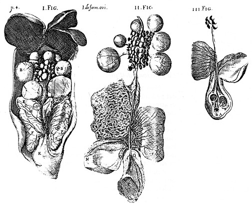
Figurarum
uterorum Gallinae in quibus ovum generatur, |
Caption
of the images of the uteri of the hen |
|
|
|
|
Primae Figurae. |
First
figure |
|
Secundae Figurae. |
Second
figure |
|
Nota
quod non apparet infundibulum quippe facile dissecando dilaceratur
debet inter ovarium, et secundum uterum adnotari. |
Note
that the infundibulum doesn't appear because it is easily torn by the
dissection and it must be placed between the ovary and the second
uterus. |
|
Figurae
Podicis explicatio. |
Explanation
of the figure of the cloaca. |
|
Tertiae Figurae. |
Third
figure |
![]()
[1] Aristotele Historia animalium I 13, p. 505b 1-2.
[2] Per esempio i Miriapodi (Myriapoda = 10.000 piedi) sono una superclasse di artropodi dall'elevato numero di zampe suddivisi in Millepiedi e Centopiedi.
[3] Aorta e vena cava caudale. Vedere questa pagina di Summa Gallicana: www.summagallicana.it/Volume3/C.VIII.a.htm.
[4] Aristotele Historia animalium VI 2, 559b 6 sqq..
[5] Probabilmente č il setto postepatico, plica peritoneale diretta dal fegato alla parete posteriore della cavitŕ addominale.
[6] Aristotele Historia animalium VI 10, 565a 8 sqq. dove 'cintura' corrisponde al greco ὑπόζωμα = diaframma.
[7] Citazione non identificabile.
[8] Aristotele forse De generatione animalium III 2, 752b 3 (dove il paragone č con un flauto, αὐλός).
[9] Aristotele De generatione animalium III 2, p. 752b 6.
[10] Aristotele Historia animalium VI 2, 752a 11 sqq.
[11] L'italiano calaza deriva dal greco chálaza, grandine, per l'aspetto particolare dei cordoncini che nell'uovo privato di guscio ricordano due chicchi di grandine; chálaza č derivato a sua volta da una radice indeuropea che significa ghiaccio. Le calaze si dipartono da ciascun polo della cellula uovo e sono dirette secondo l’asse maggiore del guscio. Si tratta di strutture cordoniformi avvolte su se stesse. Verso il polo ottuso si dirige una sola calaza, mentre dall'altro lato ne esistono due tra loro intimamente ritorte. Originano a livello dello strato calazifero e terminano da ciascun lato nella regione dei legamenti dell'albume.
[12] Si tratta dell'apertura della Borsa di Fabrizio o Timo cloacale. § Secondo Fabrizi, ciň che oggi č un organo linfatico, era invece una borsa in cui finivano il pene e gli spermatozoi del gallo. Si vede che analizzň solamente la cloaca delle galline. Infatti la borsa č presente anche nel gallo, e non solo nel gallo che per motivi contingenti viene montato da altri galli. Nella gallina gli spermatozoi del gallo trovano accoglienza molto piů in alto, e precisamente 50-80 cm dallo sbocco dell'ovidutto in cloaca: si tratta delle fossette ghiandolari, dove vengono immagazzinati. Le fossette ghiandolari si trovano nel punto di giunzione dell'infundibolo con il magnum.
[13] Si emenda in base alla nota contenuta a pagina 229 della traduzione di Howard Adelmann (The formation of the egg and of the chick - Ithaca NY, Cornell University Press, 1942).