Fabripullus
The Chick of Girolamo Fabrizi
The iconography of the chick
The
asterisk * indicates that the item is present in lexicon ![]()
|
Figurarum |
Explanation
of the figures |
|
|
|
|
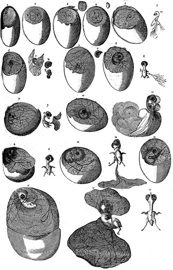
[62] Primae
figurae Ovi secundi diei ab incubatione. |
figure
1 – of an egg on 2nd day of incubation. |
|
2. Figurae 3. ovi. |
figure
2 – of an egg on 3rd day |
|
3. Figurae 4. ovi. |
figure
3 – of an egg on 4th day |
|
4. Figurae 4.
ovi. |
figure
4 – of an egg on 4th day |
|
5. Figurae 5. ovi. |
figure
5 – of an egg on 5th day |
|
6. Figurae 6. ovi. |
figure
6 – of an egg on 6th day |
|
7. Figurae 7. ovi. |
figure
7 – of an egg on 7th day |
|
8. Figurae 8. ovi. |
figure
8 – of an egg on 8th day |
|
9. Figurae 9. ovi. |
figure
9 – of an egg on 9th day |
|
10. Figurae 10. ovi. |
figure
10 – of an egg on 10th day |
|
11. Figurae 11. ovi. |
figure
11 – of an egg on 11th day |
|
12. Figurae 12. ovi. |
figure
12 – of an egg on 12th day |
|
13. Figurae 13. ovi. |
figure
13 – of an egg on 13th day |
|
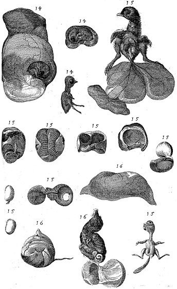
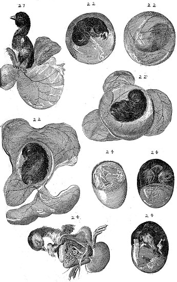
Quatuor
sequentes Figurae quae indici praeponi debuerant, exhibent
quotidianas in formatione pulli perfectiones et observationes, a
decimoquarto die usque ad vigesimumquartum, idest usque ad
exclusionis tempus. Numeri ergo singulis figuris additi diem
designant, a tempore incubationis. Partium vero pulli distinctio
facile colligitur ex tabula indici praefixa; ut minime necessarium
fuerit ea causa insculpere figuris notas distinctarum partium
indices. |
The
following four figures, which should have been placed before the
index, show the improvements and the observations of every day about
the formation of the chick starting from 14th until 24th day, that
is, until the time of hatching. Thence the numbers added to each
figure designate the day starting from the beginning of the
incubation. The distinction of the parts of the chick is easily
inferred from the table placed before the index, so that it was
quite unnecessary to add to the figures the indicative notes of the
single parts. |
|
The
egg after 14-15-16 days of incubation |
|
|
|
|
|
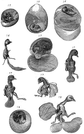
The
egg after 15-16-17-18 days of incubation |
|
|
|
|
|
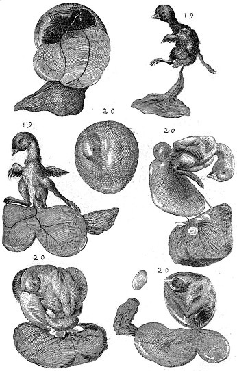
The
egg after 19-20 days of incubation |
|
|
|
|
|
The
egg after 21-22-23-24 days of incubation |
|
|
|
|
![]()
[1] Daotto o daoto o real da 8 (noto anche come pezzo da otto, dollaro spagnolo, real de a ocho, o in inglese eight real coin, Spanish dollar) č una moneta d'argento, dal valore di otto real, che fu coniata nell'Impero spagnolo dopo una riforma della monetazione nel 1497. Ebbe corso legale negli Stati Uniti fino all'emissione dell'Act del Congresso che abolě la pratica nel 1857. A causa dell'ampia diffusione in Europa, nelle Americhe e nell'Estremo Oriente, divenne la prima moneta mondiale verso la fine del XVIII secolo.