Lessico
Apofisi
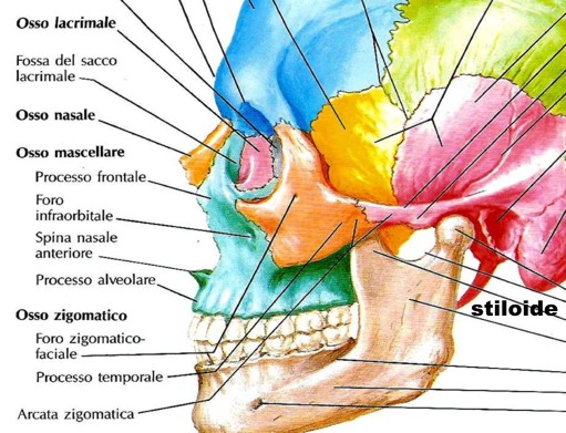
stiloide
della rocca petrosa del cranio
![]()
Stiloide viene dal greco stűlos, colonna, pilastro. In anatomia con l’aggettivo stiloide si indicano le apofisi ossee a forma di sottile colonna, appuntita all'estremitŕ. La piů importante č l'apofisi o processo stiloide dell'osso temporale, situato sulla faccia posteriore della rocca petrosa, punto di inserzione di numerosi muscoli e legamenti stiloidei.
Altri due processi stiloidei sono quelli dell’ulna e del radio situati in corrispondenza dell’articolazione del polso. Il processo stiloide che somiglia maggiormente allo sperone del gallo - o plęktron - č l’apofisi stiloide della rocca petrosa.
Aristotele![]() usa il
vocabolo plęktron per indicare un componente della caviglia, con ogni
probabilitŕ il malleolo laterale, corrispondente alla porzione distale
dell’osso che va sotto il nome di perone – o fibula – che a livello del
malleolo somiglia alla punta di una lancia, detta anch’essa plęktron, la cui etimologia viene dal verbo plęssř che significa
colpire, ferire. Anche il malleolo interno – o tibiale – ha forma
appuntita, ma si tratta di un plęktron
di dimensioni minori rispetto a quelle del perone.
usa il
vocabolo plęktron per indicare un componente della caviglia, con ogni
probabilitŕ il malleolo laterale, corrispondente alla porzione distale
dell’osso che va sotto il nome di perone – o fibula – che a livello del
malleolo somiglia alla punta di una lancia, detta anch’essa plęktron, la cui etimologia viene dal verbo plęssř che significa
colpire, ferire. Anche il malleolo interno – o tibiale – ha forma
appuntita, ma si tratta di un plęktron
di dimensioni minori rispetto a quelle del perone.
Ricordo che a una lezione di anatomia al 2° anno del mio corso di laurea in Medicina presso l'Universitŕ di Pavia (1962), una mia compagna di corso chiede al docente Luigi Cattaneo: «Scusi, Professore, si dice perňne o pérone?» - Cattaneo subito risponde: «Mi scusi, sa, ma si dice cogliňne o cňglione?» – Infatti č giusto perňne: dal greco femminile perónë = spillone, fibbia, fermaglio.
Osso temporale
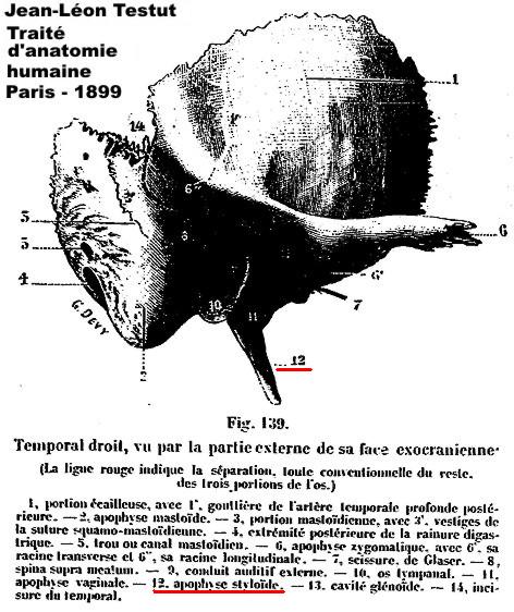
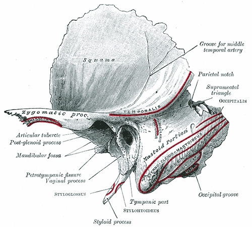
L'osso temporale č un osso pari e simmetrico del cranio, di cui occupa la parte laterale e inferiore. Nella sua compagine trovano sede l'organo dell'udito e l'apparato vestibolare e corrono importanti formazioni vascolari e nervose (carotidi, nervi acustico e facciale, ecc.). Ha forma assai irregolare ed č grossolanamente simile a una valva di conchiglia. Viene distinto in tre regioni: la porzione superiore o squama, laminare, espansa, disposta sagittalmente, da cui si stacca l'apofisi zigomatica, che saldandosi con l'osso zigomatico forma l'omonima arcata; la porzione timpanica, che insieme a un tratto della squama e alla parte petrosa delimita la cavitŕ timpanica; la porzione periotica o petromastoide che presenta inferiormente il processo stiloideo o stiloide, a sua volta suddivisa in una parte petrosa o piramide, entro cui si trova la cavitŕ dell'orecchio interno, e in una mastoidea, in rapporto con la parte postero-inferiore della precedente, che termina con il processo mastoideo.
Petrous
portion
of the temporal bone
The petrous portion of the temporal bone or pyramid is pyramidal and is wedged in at the base of the skull between the sphenoid and occipital. Directed medialward, forward, and a little upward, it presents for examination a base, an apex, three surfaces, and three angles, and contains, in its interior, the essential parts of the organ of hearing.
The base is fused with the internal surfaces of the squama and mastoid portion. The apex, rough and uneven, is received into the angular interval between the posterior border of the great wing of the sphenoid and the basilar part of the occipital; it presents the anterior or internal orifice of the carotid canal, and forms the postero-lateral boundary of the foramen lacerum.
The anterior surface forms the posterior part of the middle fossa of the base of the skull, and is continuous with the inner surface of the squamous portion, to which it is united by the petrosquamous suture, remains of which are distinct even at a late period of life. It is marked by depressions for the convolutions of the brain, and presents six points for examination:
1 -
near the center, the arcuate eminence (eminentia arcuata) which indicates the
situation of the superior semicircular canal.
2 - in front of and a little lateral to this eminence, a depression indicating
the position of the tympanic cavity: here the layer of bone which separates
the tympanic from the cranial cavity is extremely thin, and is known as the
tegmen tympani
3 - a shallow groove, sometimes double, leading lateralward and backward to an
oblique opening, the hiatus of the facial canal, for the passage of the
greater superficial petrosal nerve and the petrosal branch of the middle
meningeal artery
4 - lateral to the hiatus, a smaller opening, occasionally seen, for the
passage of the lesser superficial petrosal nerve
5 - near the apex of the bone, the termination of the carotid canal, the wall
of which in this situation is deficient in front
6 - above this canal the shallow trigeminal impression for the reception of
the semilunar ganglion.
The posterior surface forms the front part of the posterior fossa of the base of the skull, and is continuous with the inner surface of the mastoid portion. Near the center is a large orifice, the internal acoustic meatus, the size of which varies considerably; its margins are smooth and rounded, and it leads into a short canal, about 1 cm. in length, which runs lateralward. It transmits the facial and acoustic nerves and the internal auditory branch of the basilar artery.
The lateral end of the canal is closed by a vertical plate, which is divided by a horizontal crest, the crista falciformis, into two unequal portions. Each portion is further subdivided by a vertical ridge into an anterior and a posterior part.
In the portion beneath the crista falciformis are three sets of foramina; these openings together with this central canal transmit the nerves to the cochlea:
- one
group, just below the posterior part of the crest, situated in the area
cribrosa media, consists of several small openings for the nerves to the
saccule;
- below and behind this area is the foramen singulare, or opening for the
nerve to the posterior semicircular duct;
- in front of and below the first is the tractus spiralis foraminosus,
consisting of a number of small spirally arranged openings, which encircle the
canalis centralis cochleć.
The portion above the crista falciformis presents behind, the area cribrosa superior, pierced by a series of small openings, for the passage of the nerves to the utricle and the superior and lateral semicircular ducts, and, in front, the area facians, with one large opening, the commencement of the canal for the facial nerve (aqućductus Fallopii).
Behind the internal acoustic meatus is a small slit almost hidden by a thin plate of bone, leading to a canal, the aqućductus vestibuli, which transmits the ductus endolymphaticus together with a small artery and vein.
Above and between these two openings is an irregular depression which lodges a process of the dura mater and transmits a small vein; in the infant this depression is represented by a large fossa, the subarcuate fossa, which extends backward as a blind tunnel under the superior semicircular canal.
1 -
Crista falciformis.
2 - Area facialis, with (2’) internal opening of the facial canal.
3 - Ridge separating the area facialis from the area cribrosa superior.
4 - Area cribrosa superior, with (4’) openings for nerve filaments.
5 - Anterior inferior cribriform area, with (5’) the tractus spiralis
foraminosus, and (5’’) the canalis centralis of the cochlea.
6 - Ridge separating the tractus spiralis foraminosus from the area cribrosa
media.
7 - Area cribrosa media, with (7’) orifices for nerves to saccule.
8 - Foramen singulare.
The inferior surface is rough and irregular, and forms part of the exterior of the base of the skull. It presents eleven points for examination:
1 -
near the apex is a rough surface, quadrilateral in form, which serves partly
for the attachment of the Levator veli palatini and the cartilaginous portion
of the auditory tube, and partly for connection with the basilar part of the
occipital bone through the intervention of some dense fibrous tissue;
2 - behind this is the large circular aperture of the carotid canal, which
ascends at first vertically, and then, making a bend, runs horizontally
forward and medialward; it transmits into the cranium the internal carotid
artery, and the carotid plexus of nerves;
3 - medial to the opening for the carotid canal and close to its posterior
border, in front of the jugular fossa, is a triangular depression; at the apex
of this is a small opening, the aqućductus cochleć, which lodges a tubular
prolongation of the dura mater establishing a communication between the
perilymphatic space and the subarachnoid space, and transmits a vein from the
cochlea to join the internal jugular;
4 - behind these openings is a deep depression, the jugular fossa, of variable
depth and size in different skulls; it lodges the bulb of the internal jugular
vein;
5 - in the bony ridge dividing the carotid canal from the jugular fossa is the
small inferior tympanic canaliculus for the passage of the tympanic branch of
the glossopharyngeal nerve;
6 - in the lateral part of the jugular fossa is the mastoid canaliculus for
the entrance of the auricular branch of the vagus nerve;
7 - behind the jugular fossa is a quadrilateral area, the jugular surface,
covered with cartilage in the fresh state, and articulating with the jugular
process of the occipital bone;
8 - extending backward from the carotid canal is the vaginal process, a
sheath-like plate of bone, which divides behind into two laminć; the lateral
lamina is continuous with the tympanic part of the bone, the medial with the
lateral margin of the jugular surface;
9 - between these laminć is the styloid process, a sharp spine, about 2.5 cm.
in length;
10 - between the styloid and mastoid processes is the stylomastoid foramen; it
is the termination of the facial canal, and transmits the facial nerve and
stylomastoid artery;
11 - situated between the tympanic portion and the mastoid process is the
tympanomastoid fissure, for the exit of the auricular branch of the vagus
nerve.
The superior angle, the longest, is grooved for the superior petrosal sinus, and gives attachment to the tentorium cerebelli; at its medial extremity is a notch, in which the trigeminal nerve lies.
The posterior angle is intermediate in length between the superior and the anterior. Its medial half is marked by a sulcus, which forms, with a corresponding sulcus on the occipital bone, the channel for the inferior petrosal sinus. Its lateral half presents an excavation — the jugular fossa — which, with the jugular notch on the occipital, forms the jugular foramen; an eminence occasionally projects from the center of the fossa, and divides the foramen into two.
The anterior angle is divided into two parts—a lateral joined to the squama by a suture (petrosquamous), the remains of which are more or less distinct; a medial, free, which articulates with the spinous process of the sphenoid.
At the angle of junction of the petrous part and the squama are two canals, one above the other, and separated by a thin plate of bone, the septum canalis musculotubarii (processus cochleariformis); both canals lead into the tympanic cavity.
- The
upper one (semicanalis m. tensoris tympani) transmits the Tensor tympani.
- The lower one (semicanalis tubae auditivae) forms the bony part of the
auditory tube.
The tympanic cavity, auditory ossicles, and internal ear, are described with the organ of hearing.
![]()